QUIP-RS
Drosophila embryonic salivary gland and germ cell migration
QUIP-RS
Drosophila embryonic salivary gland and germ cell migration
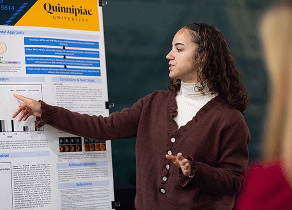
Hailey Tolson ’25, performed this study to analyze G-coupled-proteins in fruit flies to better understand their roles and functions during cell migration in humans.
Overview
This study looked closely at G-protein-coupled receptors (GPCRs) by observing their role in cell migration in fruit flies. This study aimed to identify the role GPCRs have in humans and how they influence the embryonic migratory paths of germ cells and salivary glands.
Researcher
Hailey Tolson '25
Biology
College of Arts & Sciences
Drosophila embryonic salivary gland and germ cell migration is not influenced by GPCRs mthl8 and CG15614
Abstract
G-protein-coupled receptors (GPCRs) are a seven-pass transmembrane protein superfamily conserved across several taxa. They are crucial for signal transduction to induce cellular and physiologic responses (Hanlon and Andrew 2015). Over 800 GPCRs are encoded in humans (Fredriksson and Schiöth 2005); this abundance makes them a crucial drug target for treating conditions such as hypotension with a-2 adrenergic agonists, or ß-blockers to treat arrhythmias. Nonetheless, the exact function of many GPCRs remains elusive in humans, presenting a hindrance to the development of novel therapies. Here, we examine GPCRs in the context of cell migration in fruit flies to better understand their role in cell migration.
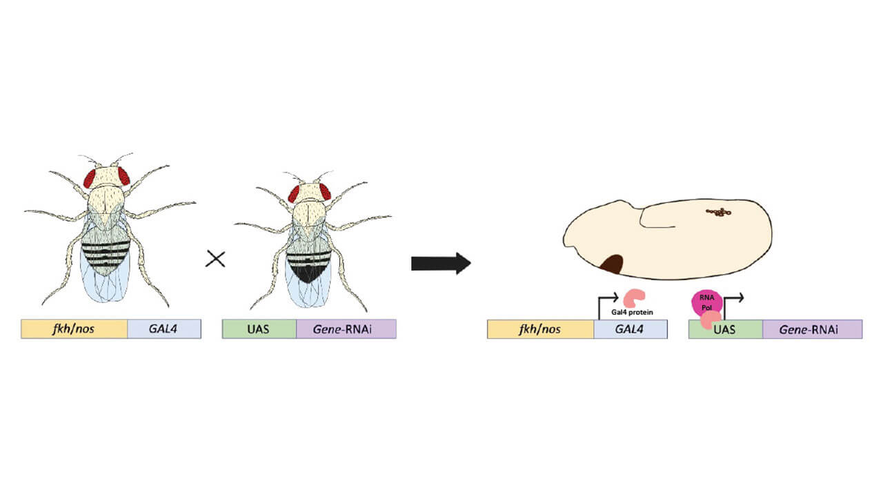
There are 116 GPCRs in the fly genome divided into four families and thirteen subfamilies, making fruit flies an excellent, simplified model to study GPCRs. Despite the wide usage of Drosophila as a model organism, GPCRs have been understudied in embryonic development and tissue migration. The Drosophila salivary glands (SG) are specified as two 140 cell placodes on the ventral surface of the embryo that invaginate dorsally and migrate posteriorly as an intact tissue through six stages of development. Germ cells (GC) migrate as individual cell migration from the midgut of the embryo to the eventual gonad. In this study, we characterized two GPCRs in embryonic SG and GC migration: methuselah-like 8 (mthl8), expressed in the optic lobe, and CG15614, whose expression pattern is largely unknown (Gramates et al. 2022). GPCR knockdown (KD) was targeted to the SGs with a forkhead promoter or to the GCs with nanos using the UAS-Gal4 system paired with DRSC/TRiP RNAi fly lines (Hu et al. 2021).
KD embryos were collected and underwent immunohistochemistry with CrebA and Vasa to stain for either SGs or GCs, respectively, before microscopic imaging. Mismigration of GCs or SGs in either the mthl8 KD or CG15614 KD did not differ significantly from wildtype tissue mismigration. Therefore, these GPCRs are unlikely to play a major role in embryonic SG or GC migration.
Background
Cell migration refers to the intentional movement of cells and tissues for their future functionality (Kolesnikov and Beckendorf 2005; Fig 1A).
The overall goal of the Hanlon lab is to characterize all 116 fruit fly GPCRs in SG and GC migration
Mthl8 is a secretin-like GPCR in the Methuselah subfamily (Hanlon and Andrew 2015; Gramates et al. 2022) with high embryonic expression (Fig 2A) and moderate single-cell RNA levels in tendon and interneuronal cells and low expression in germline cells.
CG15614 is an unclassified GPCR with unknown functions within the Rhodopsin-like family (Hanlon and Andrew 2015; Gramates et al. 2022), with low embryonic expression (Fig 2B) and moderate expression in interneuronal and female germline cells.
Hypothesis
mthl8 was not hypothesized to affect embryonic SG or GC migration due to its predominant expression in the embryonic optic lobe of Drosophila. CG15614 was not hypothesized to affect embryonic SG or GC migration per its limited expression data to neurons, epithelial cells, and Malpighian tubules.
Experimental Approach
- Knockdown GPCRs with TRiP RNAi fly lines TRiP.HMC04996 (mthl8) and TRiP.GL01053 (CG15614)
- Collect RNAi embryos, dechorinate in bleach, fix in formaldehyde solution, and devitellinize in methanol and store at 4.C
- Incubate with primary RbαVasa or RbαCrebA, then secondary RtαRb biotin, develop the stain with Vectastain and DAB substrate kits, and store in methyl salicylate at 4.C
- Image stained embryos with Jenoptic GRYPHAX microscope photo software, stage and score embryos, then analyze data in Microsoft Excel.
Conclusions and Next Steps
Conclusions
- mthl8 was hypothesized to not affect SG migration.
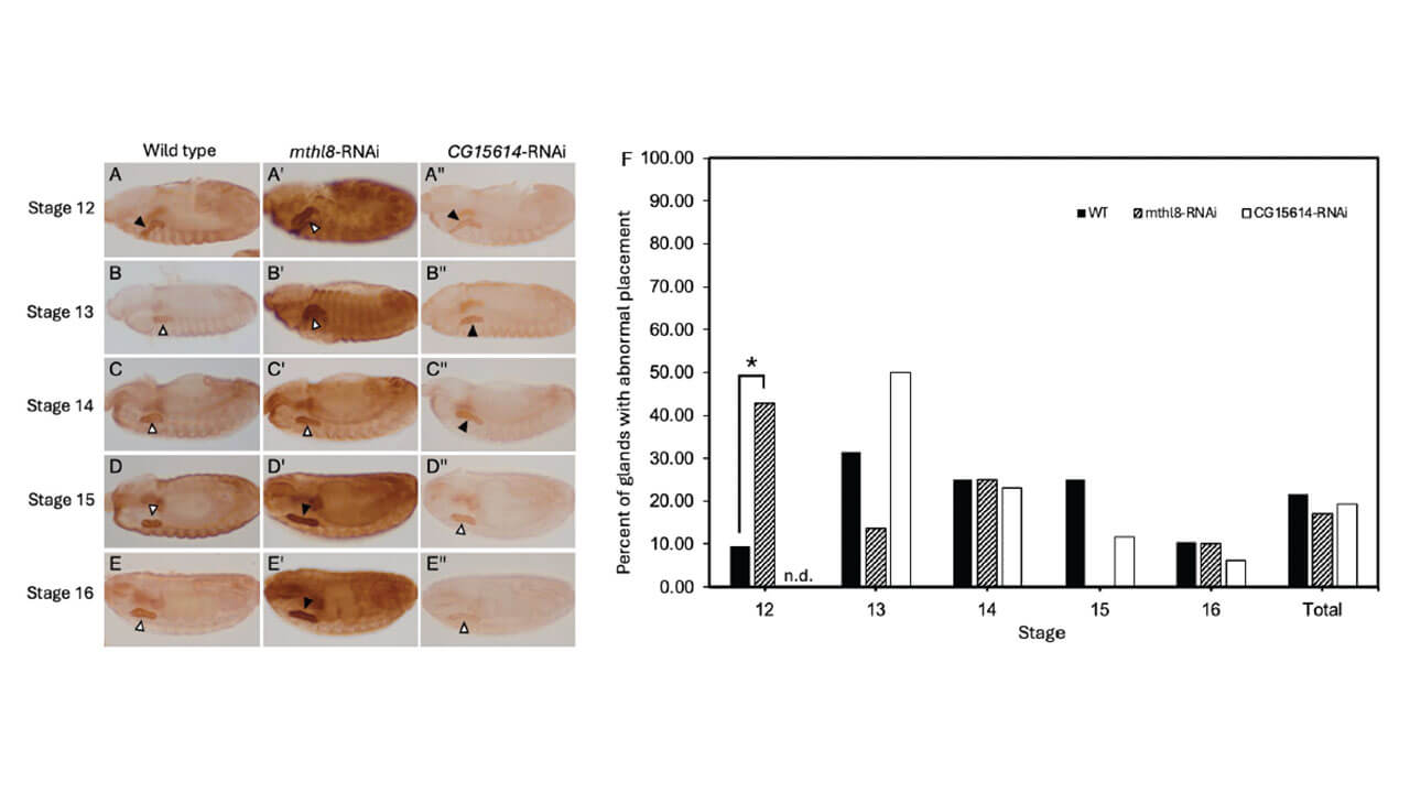
- Stage 12 mthl8-RNAi embryos had a significantly higher percentage of SGs with abnormal placement than WT, but stages 13-16 and the total percentages were not statistically significant (Fig. 4F). Stage 12 SG mismigration in mthl8-RNAi embryos is likely stochastic. We do not yet have evidence to say whether mthl8 is involved in GC migration.
- The second GPCR of interest, CG15614, was also hypothesized to not affect SG or GC migration.
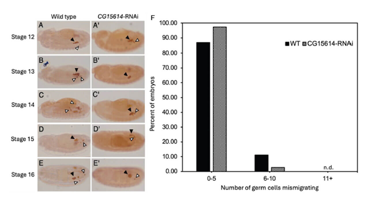
- This hypothesis is supported by the data, as the likelihood of CG15614- RNAi GCs mismigrating is not statistically different than WT GCs mismigrating (Fig. 5F).
- More data is needed to determine strong effects in SG migration, but initial results suggest it is not an important factor (Fig. 5).
Future Directions
- Tre1 is known to affect germ cell migration cell autonomously, but published data suggest that loss of Tre1 may also affect the shape of the midgut pocket (Fig. 5E and F, adapted from Lin et al. 2020).
- We propose to examine the shape of the midgut pocket in Tre1 and gcl mutants to better understand how the presence of Tre1 and germ cells impact midgut pocket shape, respectively.
Acknowledgements
We are incredibly grateful to the QUIP-RS committee for selecting our project to receive funding in this competitive program as the Hanlon lab continues to profile GPCRs’ role in embryonic cell migration. We also extend our gratitude to the Bob MHS '84 and Carlita Gasparini Fund for financial support to present our work at national conferences and providing more opportunities for 3+1 Biology, Biochemistry, and MCB students.
References
Fredriksson, R. and Schiöth, H. B. 2005. The Repertoire of G-Protein-Coupled Receptors in Fully Sequenced Genomes. Mol. Pharm. 67(5): 1414-1425. 10.1124/mol.104.009001
Gramates, L. S., Agapite, J., Attrill, H., Calvi, B. R., Crosby, M. A., Dos Santos, G., Goodman, J. L., Goutte-Gattat, D., Jenkins, V. K., Kaufman, T., Larkin, A., Matthews, B. B., Millburn, G., Strelets, V. B. 2022. FlyBase: a guided tour of highlighted features. Genetics 220(4). iyac035.
Hanlon, C. D. and Andrew, D. J. 2015. Outside-in signaling – a brief review of GPCR signaling with a focus on the Drosophila GPCR family. J. Cell Sci. 128(19): 3533-3542. 10.1242/jcs.175158
Hu Y, Comjean A, Rodiger J, Liu Y, Gao Y, Chung V, Zirin J, Perrimon N, Mohr SE. FlyRNAi.org-the database of the Drosophila RNAi screening center and transgenic RNAi project: 2021 update. Nucleic Acids Res. 2021 Jan 8;49(D1):D908-D915. doi: 10.1093/nar/gkaa936. PMID: 33104800; PMCID: PMC7778949.
Kolesnikov, T. and Beckendorf, S. K. 2005. NETRIN and SLIT guide salivary gland migration. Dev. Bio. 284(1): 102-111. 10.1016/j.ydbio.2005.04.037
Ribeiro, C., Petit, V., Affolter, M. 2003.
Signaling systems, guided cell migration, and organogenesis: insights from genetic studies in Drosophila. Dev. Bio. 260(1): 1-8. doi.org/10.1016/S0012-1606(03)00211-2
Warrington, S. J., Strutt, H., Strutt, D. 2013. The Frizzled-dependent planar polarity pathway locally promotes E-cadherin turnover via recruitment of RhoGEF2. Development. 140(5): 1045-1054. http://doi.org/10.1242/dev.088724
Williamson, A. and Lehmann, R. 1996. Germ Cell Development in Drosophila. Annu. Rev. Cell Dev. Biol. 12: 365-391. 10.1146/annurev.cellbio.12.1.365
Results and Conclusions
Professional Application
”I was looking for research experiences to better prepare me for both medical school and a job in the research industry. When I heard about QUIP-RS, I knew it was a perfect opportunity to expand on this interest, and even better with someone I’m close to as my mentor. I gained a lot more insight into the field of scientific research than I was anticipating and fell in love with the process more than I thought; I’ve gone from an aspiring MD to an aspiring MD/PhD after this program! There is still much to learn, but I’m so grateful this was my first real taste of research.“ - Hailey Tolson ’25
Faculty Mentor
For Further Discussion
This serves as an overview of the project and does not include the complete work. To further discuss this project, please email Hailey Tolson.
Quinnipiac University Interdisciplinary Program for Research and Scholarship
Open to students of all majors, QUIP-RS provides up to $5,000 in funding for undergraduate students to conduct research or complete creative projects alongside faculty mentors. This intensive 8-week program enables students to develop scholarly skills while encouraging discussion about successes and shortcomings with fellows and mentors.
Explore Our Areas of Interest
We've sorted each of our undergraduate, graduate and doctoral programs into unique Areas of Interest. Explore these categories to discover which programs and delivery methods best align with your educational and career goals.